Autoimmune hemolytic anemia (AIHA) is a type of hemolytic anemia caused by the production of anti-self-erythrocyte antibodies due to immune dysfunction, binding to red blood cell surface antigens, or activating complement to accelerate the destruction of red blood cells. According to reports, the incidence of AIHA in adults is 0.08‰~0.3‰ per year, the prevalence is 0.17‰, and the mortality rate is 11.0%. According to the cause of the disease, it can be divided into primary and secondary. Primary AIHA often has no underlying disease and the cause of the disease is unclear; secondary AIHA is associated with lymphatic system malignancies and immune-related diseases, such as lymphoma, chronic lymphocytic leukemia, ulcerative colitis, systemic lupus erythematosus, certain drugs and bacterial and viral infections. Current studies on the cause of AIHA have shown that it is mostly related to T lymphocytes and B lymphocytes. However, as scientific research gradually deepens, it is discovered that diseases are also accompanied by gene and protein abnormalities. Regulating these abnormal genes and target proteins may become an important way to treat such diseases in the future.

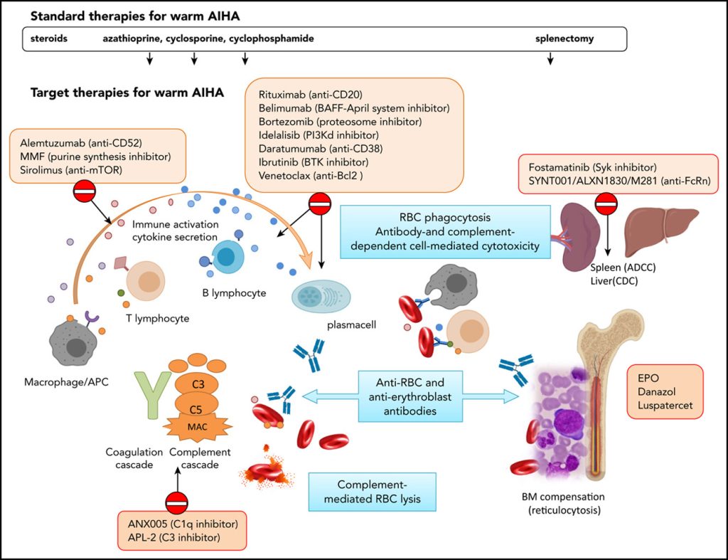
Figure 1. Treatment of febrile autoimmune hemolytic anemia.
Primary AIHA
Genetic Level
Copper/zinc superoxide dismutase genes (SOD1 genes): Superoxide dismutase (SOD) is an active substance derived from living organisms that can eliminate harmful substances produced by organisms during metabolism. Continuous supplementation of SOD in the human body has a special anti-aging effect. Studies have shown that SOD is associated with a variety of autoimmune diseases, such as autoimmune cerebrospinal meningitis and rheumatoid arthritis. SOD1 is a subtype of SOD dimer containing Cu and Zn atoms. To date, most studies on the SOD1 gene have focused on changes in the relevance of amyotrophic lateral sclerosis. However, recent studies have found that oxidative stress deficiency of SOD1 can also lead to anemia and autoimmune reactions. NZB mice are mice with autoimmune diseases that will spontaneously develop AIHA after 4 to 6 months. The study found that as the course of the disease in NZB mice prolonged, the levels of methemoglobin and lipid peroxidation products in the body continued to increase, and consistent with this, the level of reactive oxygen species (ROS) in erythrocytes also continued to increase, and was positively correlated with the degree of anemia and the incidence of AIHA. However, the level of ROS in erythrocytes in hSOD1-Tg/NZB mice carrying the human SOD1 gene decreased significantly. The SOD1 gene reduces the incidence of AIHA by inhibiting the level of ROS in erythrocytes.
Protein Level
Human erythrocyte anion exchanger 1 (AE1): Human erythrocyte anion exchanger 1 is located in the third band in erythrocyte membrane protein electrophoresis, also known as band 3 protein. It is encoded by the SLC4A1 gene and is one of the most important proteins on the erythrocyte membrane. Band 3 protein is not only the main attachment point of the membrane skeleton, but also participates in the transmembrane transmission and functional regulation of various substances and information. Any changes in its structure or content may affect the integrity of the erythrocyte membrane structure and the response deformation ability of the entire cell. Membrane protein band 3 regulates the interaction with other cytoskeleton proteins and key enzymes of metabolic pathways, and mediates the phosphorylation of Lyn kinase and Src family kinase, thereby jointly maintaining the overall balance of erythrocytes. For example, streptolysin can inhibit the activity of band 3 protein, allowing chloride ions to quickly enter erythrocytes, destroying the stability of the cell membrane, and causing hemolysis. In addition, a series of serological and immunohistochemical tests were performed on 120 AIHA patients, and it was found that warm anti-self IgG antibodies can specifically act on band 3 protein, and it was also proved that it is an important target of warm antibodies in AIHA patients. Therefore, band 3 protein plays a vital role in the destruction of red blood cells and antibodies. Regulating the function of band 3 protein may reduce the hemolysis of AIHA and thus alleviate the disease process.
Stromal interaction molecule (STIM): store-operated calcium ion channel (SOCE) is one of the important channels that mediate the entry of extracellular calcium ions into cells. Its core protein is composed of the stromal interaction molecule (STIM) located on the endoplasmic reticulum and the calcium release-activated calcium channel modulator (ORAI) located on the cell membrane. At present, studies have found that STIM protein is divided into two subtypes, STIM1 and STIM2, and their main functions are slightly different. In the process of the body fighting against pathogens, STIM protein can change due to the increase and decrease of calcium ions in the cell, accumulate and translocate to the vicinity of the plasma membrane. The accumulated STIM protein will then activate the Orai1 protein, prompting the calcium release activated calcium channel (CRAC) to open. The influx of calcium ions leads to cell activation, thereby activating immune cells. STIM actually has the dual functions of regulating the opening and selectivity of CRAC channels. If the STIM1 and STIM2 proteins of T lymphocytes are deficient, autoimmune diseases of the exocrine glands, such as Sjögren’s syndrome, will occur. Researchers have found that STIM2 is closely related to cell migration and cytokine changes downstream of G protein-coupled receptors and TLR4 activation in macrophages. Like STIM1, it can cause the release of stored calcium and phagocytosis. In addition, in the AIHA model, STIM1 also activates C5aR-mediated FCγR, thereby causing the occurrence and development of the disease. Therefore, STIM1 and STIM2 act jointly and independently on macrophages in the fatal hemolysis process. After blocking the STIM protein-related functions, the mortality rate of AIHA model mice and LPS-induced septic mice was significantly reduced, further indicating that the inhibition of STIM protein may help alleviate inflammatory and autoimmune diseases.
Secondary AIHA
Gene Level
Immunoglobulin variable heavy chain region (IGVH) gene: The immunoglobulin variable heavy chain region is currently an important molecular indicator for predicting the condition and prognosis of chronic lymphocytic leukemia. It is generally believed that patients with somatic hypermutation of the IGHV gene sequence have a better prognosis and a longer overall survival than those without mutations. In a retrospective study of 585 Chronic lymphocytic leukemia (CLL) patients, it was found that the non-mutation state of IGHV was an independent influencing factor for the secondary occurrence of AIHA in CLL patients, and the median survival of AIHA disease development in non-mutated patients was significantly shortened. It is speculated that it may play an important role in specific B cell receptor subsets, but the specific molecular mechanism of action is still unclear.
MicroRNA: MicroRNA is a type of non-coding single-stranded RNA molecule with a length of about 22 nucleotides encoded by endogenous genes, which participates in post-transcriptional gene expression regulation in animals and plants. It has been found that microRNA is differentially expressed in many immune diseases. Therefore, drugs that act on abnormal microRNAs may be a new research direction to achieve therapeutic effects, such as systemic lupus erythematosus, chronic idiopathic urticaria, etc. In the process of studying the physiological and pathological mechanisms of AIHA secondary to CLL, it was found that a total of 9 miRNAs (miR-19a, miR-20a, miR-29c, miR-146b-5p, mir-186, miR-223, mir-324-3p, mir-484miR-660) were downregulated in expression. Two of the miRNAs (miR-20a and miR-146b-5p) are involved in autoimmune phenomena, especially miR-146b-5p, which is involved in both autoimmune diseases and chronic lymphocytic leukemia. Further research on this microRNA found that miR-146b-5p is closely related to regulating CD80, which is a molecule closely related to the synapse between B lymphocytes and T lymphocytes and the restoration of cell antigen presentation ability. Therefore, research on microRNA may become a new hotspot for the treatment of related immune diseases in the future.
Protein Level
B cell adsorption factor 1 (BCA-1): B cell adsorption factor 1, also known as B cell chemoattractant (BLC) or CXCL13 (chemokineCXCligand13), is a member of the CXC chemokine family and is secreted by follicular dendritic cells and macrophages in secondary lymphoid organs. This chemokine selectively attracts B cells, including two subsets, B-1 and B-2, and exerts its effect by interacting with the chemokine receptor CXCR5. The CXCR5 receptor is currently the only known ligand, which is expressed in mature B cells, follicular helper T cells, Th17 cells, and regulatory T cells. Many studies have shown that abnormal expression of CXCL13 is associated with the development of autoimmune diseases such as rheumatoid arthritis, multiple sclerosis, and systemic lupus erythematosus. Since secondary AIHA is mostly secondary to autoimmune diseases, especially systemic lupus erythematosus. Through the case analysis of clinical AIHA patients, the researchers traced the relationship between it and systemic lupus erythematosus and found that CXCL13 and hemoglobin values were negatively correlated, while another chemokine CCl4 (chemokine (C-Cmotif) ligand4) and reticulum erythematosus values were positively correlated. They can be used as sensitive markers for the growth and decline of AIHA disease. The plasma CXCL13 level can reflect the severity of the disease, and CCl4 can be used as an indicator for determining the bone marrow hyperplasia of AIHA patients. At the same time, higher plasma soluble tumor necrosis factor receptor II levels may have a strong guiding significance for distinguishing whether it is AIHA secondary to SLE. Therefore, the use of CXCL13 antagonists may have a certain degree of delaying effect on the development of AIHA.
In short, the pathogenesis of AIHA involves the interaction of multiple factors such as genes and proteins. As the research continues to deepen, these possible influencing factors will continue to be discovered, which can provide possible drug targets for clinical treatment and provide further in-depth directions for the study of its pathogenesis.
