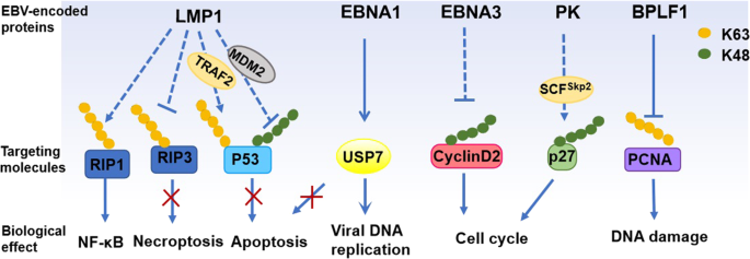
Epstein-Barr virus (EBV) is a kind of γ-herpes virus that is infected with human beings. In 1964, the Epstein team found in Burkittlymphoma (BL) that it consists of a double-chain DNA of 184 KB, mainly through saliva spread. 95%of the world’s people have been infected with EBV, which can be infected in the body and can also cause a variety of diseases and tumors. At present, tumors related to EBV infections include epithelial tumors such as nasopharyngealcarcinoma(NPC), gastric carcinoma (GC), lymphatic hematopoietic malignant tumors such as Hodgkin’s Lymphoma (HL) and Non-Hodgkin Lymphoma (NHL), BL, pyropogy-related lymphoma (PAL) and NK/T cell lymphoma and other rare tumors (such as EBV-related smooth fibroids, neuromuscular endocrine cancer). EBV’s threat to humans is far more than that. Therefore, it is particularly important to understand how EBV is infected with the host and develops in the body.

EBV Invasion Mechanism
EBV Invasion B Lymphocytes
EBV is a virus transmitted through saliva. Oral mucosal epithelial cells are the first threshold for their invasive host cells. EBV’s primary infection is considered to be caused by viruses through the oropharyngeal epithelium. Infection of naive B cells present in Waldeyer’s ring of tonsils. EBV shows obvious tendency to B lymphocytes, which is easy to infect B cells and convert initial B cells into proliferative lymphocytes. Viral glycoproteins including gp350, gHgL, gB and gp42 mediate the preferential binding of EBV to B cells by interacting with the complement receptor CR2 (CD21) on the surface of B cells, and then the envelope glycoprotein gp42 and gp85/gp25 form a fusion protein triple molecule Complex. The GP42 in the complex is combined with the HLA II molecular molecules, and caused the virus cell fusion under the participation of GP85/GP25 and GP110 glycoprotein.
EBV Invades T/NK Lymphocytes
EBV can also infect T/NK lymphocytes, but the mechanism by which EBV attaches and invades T/NK lymphocytes has not been elucidated. T/NK lymphocytes neither express CD21 nor HLA class II, but T/NK lymphocytes express some integrins, which increase when stimulated and can act as receptors for T/NK lymphocytes. Studies have shown that both primitive T lymphocytes and lymphoid progenitor cells can express CD21, and EBV can also infect primitive T cells and lymphoid progenitor cells by attaching to T lymphocytes through CD21. The double infection of T lymphocytes and NK cells in patients with chronic active EBV infection (CAEBV) has been reported in the literature, further supporting that EBV may infect the common progenitor cells of T cells and NK cells. EBV can also be transmitted from EBV-infected B cells or epithelial cells to T/NK lymphocytes by cell-to-cell infection. EBV-infected B cells can activate NK cells to acquire CD21 molecules through synaptic transfer, and the ectopic receptor leads to the binding of EBV to NK cells. Studies have shown that EBV-infected T/NK lymphocytes often express cytotoxic molecules, such as perforin, granzyme B, and T cell intracytoplasmic antigen (TIA-1). NK cells, CD8+ T cells, and γδ T cells observed in EBV-associated T/NK lymphocyte tumors are typical of EBV-infected cells and belong to the type of killer cells that attempt to kill EBV-infected B cells or epithelial cells. Cells may become infected with EBV through close contact at immune synapses.
EBV Invades Squamous Cells
EBV initially enters the body through the oropharyngeal mucosa and infects B cells through the binding of the viral envelope protein gp350 to CD21 on the surface of B cells. Epithelial cells do not express CR2, and the mechanism of how EBV invades and releases from epithelial cells is not yet clear. Studies have shown that EBV can enter tongue and pharyngeal epithelial cells through 3 pathways independent of CD21 (CR2): 1) direct cell-cell contact with EBV-infected lymphocytes through the apical cell membrane; 2) through β1 or α5β1 integrin The interaction with the EBV BMRF-2 protein mediates the entry of EBV free virus particles into the basement membrane; 3) After primary infection, EBV spreads directly across the lateral membrane to adjacent epithelial cells. The anti-EBV antigen polymer IgA can mediate the invasion of EBV into pharyngeal epithelial cells through endocytosis, and EBV bound to IgA can invade pharyngeal epithelial cells through endocytosis mediated by secretory corpuscle (SC). Elevated levels of anti-EBV-specific antigen IgA were found in mucosal secretions of NPC patients, and this EBV-IgA-SC-mediated endocytosis may represent a physiological pathway for EBV to invade nasopharyngeal epithelial cells in vivo.
EBV Invades Glandular Epithelial Cells
EBV infection of glandular epithelial cells can cause gastric cancer and bile duct cancer, and the mechanism is unclear. At least 3 models are currently speculated as the mechanism by which EBV attaches to glandular epithelial cells, which may overlap with the mechanism by which EBV invades squamous epithelial cells: 1) EBV virions with IgA specific for gp350/220 have been shown to effectively Binds to polymeric IgA receptors. Polymerized IgA is normally present in human saliva and binds to transmembrane proteins expressed on the basolateral surface of polarized epithelial cells. Internalization of the EBV-IgA-SC complex into glandular epithelial cells via an endocytic pathway is associated with an infection mechanism through the basolateral surface of the epithelial cell, possibly similar to the physiological infection of the virus in vivo. 2) It has been demonstrated that in the absence of CD21 (CR2), the gH and gL complexes can act as epithelial ligands, and EBV derived from B cells can bind to CD21 (CR2) negative epithelial cells with high affinity, but lack gH The EBV/gL complex loses its ability to bind, suggesting that the gH/gL complex present on the surface of EBV can directly bind to epithelial cell-specific receptors (such as integrins αVβ6 and αVβ8) to trigger fusion of EBV with the epithelial plasma membrane. 3) The interaction of the EBV-encoded membrane protein BMRF2 with integrins on polarized epithelial cells is another model for EBV attachment to the cell surface. The tripeptide Arg-Gly-Asp (RGD) motif in the BMRF2 molecule is presented as a ligand for rβ1, α5, α3 and αV integrins. However, BMRF2 is not a membrane protein necessary for cell-cell fusion, and very few BMRF2 molecules are present in virions. It is unclear whether the interaction of BMRF2 with integrins is primarily responsible for attachment and/or post-attachment events.
