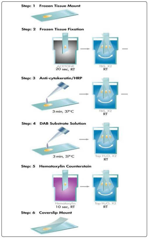
Immunohistochemistry is to apply the basic principle of immunology antigen-antibody reaction, that is, the principle of the specific binding of antigen and antibody. Through a chemical reaction, antibodies (fluorescein, enzymes, metal ions, isotopes) labeled with color reagents are developed to determine the antigens’ (peptides and proteins) localization, qualitative and relative quantitative in tissue cells.
Before using specific antibodies to detect antigens by immunohistochemistry (IHC), all potential non-specific binding sites in the tissue sample must be blocked to prevent non-specific binding of antibodies to these sites. Omitting or inadequate blocking treatment will cause antibodies or other detection reagents to bind to other unrelated sites, resulting in a higher background in the final imaging. For example, antibodies can bind to multiple types of surfaces through simple adsorption, and due to charge, hydrophobicity, and other types of interactions, they can be non-specifically adsorbed to proteins.

Generally speaking, any protein that does not specifically bind to the target antigen or antibody and other detection reagents in the IHC assay can be used as a component of the blocking solution. However, in fact, some proteins have better blocking performance than other proteins because they are easier to bind to neutral non-specific sites or have the function of stabilizing other assay components. However, there is no single protein or protein mixture that can provide the best blocking effect for all IHC experiments. Therefore, before IHC blocking, a blocking test is performed for a combination of a given specific antibody and other detection reagents, which is essential to obtain the best blocking result.
General Blocking Procedure
The blocking step of IHC is usually performed before the incubation of the primary antibody after the sample is processed. The general procedure is to incubate the processed IHC sample with an appropriate blocking buffer at ambient temperature for 30 minutes or overnight at 4°C to complete the binding of the blocking solution to the non-specific binding site. It is then necessary to perform sufficient washing after the blocking step to remove excess protein that may prevent the detection of the target antigen. However, many researchers did not wash after the blocking step because they diluted the primary antibody in the blocking buffer.
Selection of IHC Blocking Fluid
Immunohistochemistry (IHC) experiments usually include one or more blocking steps to reduce background signals and false positives. Commonly used blocking solutions mainly include: serum, protein solution and commercial buffer.
Serum Blocking
Serum is a common blocking agent because it contains antibodies that bind to non-specific sites. Among them, 1-5% (w/v) normal serum is a common blocking buffer component, because the antibody carried in the serum will bind to the reaction site and prevent the non-specific binding of the secondary antibody used in the assay. However, an important factor to consider is the use of serum from the species from which the secondary antibody is derived. This is because the serum from the first antibody species mainly binds to the reaction site, but the second antibody is the key to determining the non-specific binding antibody and the specific antibody binding to the target antigen. In addition, serum is rich in albumin and other proteins that easily bind to non-specific protein binding sites in the sample.
Protein Solution Blocking
In addition to serum, blocking buffers usually contain proteins, such as bovine serum albumin (BSA), gelatin or skimmed milk powder at a final concentration of 1-5% (w/v). Compared with the antibody concentration, the large amount or excess of these cheap and easily available proteins (alone or together) can compete with specific antibodies for non-specific sites in the sample. Many laboratories have their favorite self-made closed buffer formulas. However, it is important to ensure that this blocking buffer is free of precipitates and other contaminants that can interfere with IHC detection.
Pre-prepared Commercial Blocking Buffer
Ready-made blocking buffers can also be used to block samples in preparation for antibody processing. These buffers can contain highly purified single proteins or proprietary compounds that do not contain proteins. The advantage of using commercially available blockers is that there are many available options that have better performance than gelatin, casein or other proteins used alone, and they have a longer shelf life than homemade preparations.
Block Reminder
We provide a few important tips for your blocking experiment:
1. Try different blocking agents to select the blocking agent with the highest signal-to-noise ratio through background (negative control) and signal intensity (positive control);
2. Make sure that there is no substance in the blocking buffer that interferes with the target measurement. For example, skimmed milk powder contains biotin and is not suitable for any detection system containing biotin-binding protein.
3. Use the same blocking buffer to dilute the antibody used for the blocking step.
4. For detection based on biotin, HRP, alkaline phosphatase or the presence of endogenous chromogenic enzymes, it needs to be blocked and then tested. E.g:
- Biotin Blocking
Biotin is present in many tissues, especially in the kidney, liver and brain. By pre-incubating the tissue with avidin and then incubating with biotin to block other biotin binding on the avidin molecule Site to close it. - Endogenous Enzymes Blocking
Chromogenic detection methods usually use enzymes directly or indirectly linked to the secondary antibody to visualize antibody localization. If the enzyme is naturally present in the tissue under study, its activity must be blocked before the detection step.
Peroxidase blocking
When using HRP for detection, non-specific or high background staining may occur due to endogenous peroxidase activity. The kidney, liver and other tissues and tissues containing red blood cells (such as blood vessel tissue) all contain endogenous peroxidase. To check endogenous peroxidase activity, the tissue can be incubated with DAB substrate before the primary antibody incubation. If the tissue turns brown, endogenous peroxidase is present and a blocking step is required. An incubation in 0.3% hydrogen peroxide for 10-15 minutes is usually sufficient for blocking. - Alkaline Phosphatase(AP) Blocking
When using AP for detection, endogenous alkaline phosphatase (AP) may produce high background. It can be found in the kidneys, intestines, osteoblasts, lymphoid tissues and placenta. AP activity in frozen tissue is higher. The endogenous AP of the tissue can be detected by incubating with BCIP/NBT; if a blue color is observed, it indicates the presence of endogenous AP, so it is necessary to block it. Levamisole is used for blocking and is added with the chromogenic substrate. Before adding the primary antibody, seal the intestinal AP with a weak acid (such as 1% acetic acid). - ReduceAutofluorescence in IHC
When using fluorescent markers for detection, the tissue may auto-fluoresce, resulting in excessive background. Tissue fixation may cause autofluorescence, especially when aldehyde fixatives (such as formalin) are used, which react with amines to form fluorescent products.
Autofluorescence may also be caused by the presence of fluorescent compounds such as flavin and porphyrin. These compounds can be extracted from the tissue. However, they will remain in frozen sections that have been treated with aqueous reagents. To reduce autofluorescence, non-aldehyde fixatives (such as Carnoy’s solution) can be used, or aldehydes can be blocked by treatment with sodium borohydride or glycine/lysine. Alternatively, try using frozen tissue sections or treating the tissue with a quenching dye (for example, Bomitan Sky Blue, Sudan Black, Trypan Blue or FITC Block).
